News
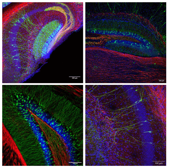
Example images of mouse brain sections following immunolabeling, tissue clearing, and confocal microscopy by students in BE-131/ES-225: Neuroengineering. Photo credit: Ananya Salem, Mira Jiang, Tomas Winegar (top, left); Samantha Sestak, Frederico Araujo, Aaron Bradford (top, right); KaiLan Mackey, Sofia Flynn, Viktor Bokisch (bottom, left and right).
Mapping the complexities of the brain is one of the greatest ambitions in neuroscience. Critical to this endeavor is the development of sophisticated imaging techniques that might provide insight into the relationship between brain structures and functions. These techniques are usually only used in highly specialized facilities, but this semester students in “BE-131/ES-225: Neuroengineering,” taught by Professor Jia Liu, Assistant Professor of Bioengineering at the Harvard John A. Paulson School of Engineering and Applied Scieces (SEAS), got hands-on experience using these advanced techniques.
Conducted in the SEAS Active Learning Labs (ALL), and led by ALL staff members Melissa Hancock and Avery Normandin, the goal of the lab was to help “students connect what they learned in lecture about scientific mechanisms and engineering principles with hands-on skills that bring these concepts to life,” said Liu. “Through tissue clearing, genetic engineering, and antibody staining, they learn how to map neuron connectivity within three-dimensional brain tissue at the single-cell level with cell-type specificity.”
“This involves a series of detailed techniques, from basic pipetting to advanced confocal microscopy, empowering students with practical experience in imaging and data analysis. These skills not only enhance their understanding of neural circuitry but also prepare them for future research in neuroengineering and brain science.”
The imaging process took over four weeks. Students began the lab with whole mouse brains, which they then sliced into thick sections. Working collaboratively in small groups, students used a technique called iDISCO labeling, which involves treating the sections with various solvents to make the brain samples optically clear. That allowed for improved resolution and greater depth of optical imaging. In collaboration with Douglas Richardson, Alex Lovely, Linda Liang, and Heather Brown-Harding from the Harvard Center for Biological Imaging (HCBI), students imaged their labeled samples with a type of microscope called a confocal laser scanning microscope.
Bioengineering concentrator Frederico Araujo, S.B. '25, appreciated the ability to experience the tissue clearing process.
“My favorite thing about the lab was the fact that we got access to a full brain research protocol – from tissue processing to data collection and analysis,” he said. “Practicing with these protocols broadens my skill set.”
Data analysis was an important element of this course following the image collection. Student groups, each imaging unique parts of the brain, generated experimental questions about what immunostaining would reveal about that particular brain region.
“The lab component of this class provided an incredibly hands-on experience, where even those of us without prior lab training could dive into a complex protocol,” said Viktor Bokisch, S.B. '26. “The format was thoughtfully designed, empowering us to achieve remarkable results: capturing stunning images of brain tissue and giving us a deep appreciation for the intricacies of neuroscience and wet lab work in general.”
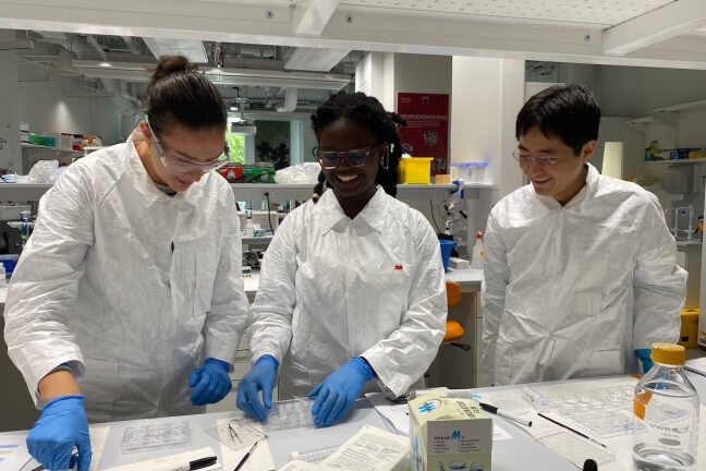
Students Kristin Otervik, Onovughakpor Otitigbe-Dangerfield, and Shikoh Hirabayashi prepare their brain sections for immunolabeling (Melissa Hancock/Active Learning Lab)
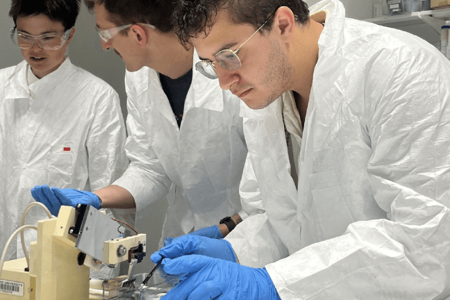
Ricardo Marrero-Alattar at the microtome sectioning a mouse brain. (Sara Elkilany)
Topics: Academics, Bioengineering
Cutting-edge science delivered direct to your inbox.
Join the Harvard SEAS mailing list.
